- Introduction
- Theory
- Application
- See also
- References
Introduction
Optical Coherence Tomography takes use of Michelson interferometer to build a 3D picture layer by layer. The photons reflected by the sample can only have interference with the reference photons when the distance between reference arm and sample arm is with the source’s coherence length. As a tomography imaging method which only records ballistic and quasiballistic photons with depth information, it is now developed to obtain optical properties including scattering coefficient of tissues and shows its reliablity and potential in non-invasive continuous blood glucose monitoring.
Theory
1. Single scattering model
According to the Beer–Lambert law, light attenuation inside tissues is exponential. The slope of this exponential attenuation is proportional to the total attenuation coefficient of ballistic photons.
[1][2]

Because  for tissues in the NIR, the exponential attenuation is proportional to the scattering coefficient. Tissue scattering properties are highly dependent on the ratio of the refractive index of scattering centers (cell membranes, cellular components, and protein aggregates),
  , to the refractive index of the interstitial fluid (ISF),
  :
:

A simplified empirical formula of Mie theory can be used to describe how  changes,[3]

Where  is radius of scattering sphere,
  is number of scattering spheres in unit volume and
  is wavelength of incident light.
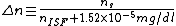
When the blood glucose concentration is increased, -- the refractive index of the ISF will be increased by  per each
  [4] due to the permeability of blood glucose.
The refractive index mismatch decrease will reduce the scattering coefficient () of tissues.
By analyzing the exponential profile of light attenuation detected by the OCT system, one can obtain information on tissue scattering properties. The principle is that the processing of OCT 2-dimensional images can get the OCT signal of light intensity as a function of depth in the logarithmic range. By linear fitting the exponential attenuation slope of light, the relative value of the tissue scattering coefficient can be obtained. Because an increase of the ISF glucose concentration in the physiological range ( ) may decrease the scattering coefficient by
  due to cell volume change[5], the glucose concentration can thus be measured.
2. Multiple scattering model based on Expansion Huygens-Fresnel principle
The multi-scattering model based on EHF theory is applicable in both homogeneous medium and non-uniform structure with multi-layers, which greatly expands the limitations of the previous method of measuring optical parameters of only shallow tissues.
In this model, the sample can be viewed as a discrete multi-layer stack. The model focuses on photon transfer at each layer boundary. This model uses the Huygens Fresnel principle to extract the extinction coefficient and anisotropy factor of each layer, so that the OCT can obtain deeper tissue information. This method is suitable for most biological tissues.
So far, the single-layer structure and the two-layer structure have been studied using this multi-scattering model, and the scattering coefficient and anisotropy factor of layer of each sample are accurately measured.[6][7][8]
Application
The OCT system is quantitatively proved to have specificity of measuring blood glucose, showing the increase of glucose concentration can decrease scattering coefficient in a linear way.[9] The high accuracy and reliability of OCT continuous monitoring of blood glucose concentration in microvasculature structure of subjects with diabetes is reported by using statistical analysis including Clarke error grid and calculation of the Pearson correlation coefficient.[10]In recently clinical trials,
OCT is reported to detect glucose through human fingertips by obtaining the optical rotation angle and the depolarization index of light.[11], and some other properties of blood such as attenuation coefficient and blood viscosity variations are obtained from OCT signals to measure blood glucose concentration.[12]
See also
- Optical coherence tomography
- Michelson interferometer
- Blood glucose monitoring
References
2. ^J. H. Lambert, Photometria sive de mensura et gradibus luminis, colorum et umbrae [Photometry, or, On the measure and gradations of light, colors, and shade] (Augsburg ("Augusta Vindelicorum"), Germany: Eberhardt Klett, 1760).
3. ^Graaff, R., Aarnoudse, J. G., Zijp, J. R., Sloot, P. M., Mul, F. F., Greve, J., & Koelink, M. H. (1992). Reduced light-scattering properties for mixtures of spherical particles: A simple approximation derived from Mie calculations. Applied Optics,31(10), 1370. doi:10.1364/ao.31.001370
4. ^Tuchin VV: Tissue Optics. Light Scattering Methods and Instruments for Medical Diagnostics. Bellingham, WA, SPIE Press, 2000
5. ^Specificity of noninvasive blood glucose sensing using optical coherence tomography technique: a pilot study
6. ^Lutomirski, R. F., & Yura, H. T. (1971). Propagation of a Finite Optical Beam in an Inhomogeneous Medium. Applied Optics,10(7), 1652. doi:10.1364/ao.10.001652
7. ^Schmitt, J. M., & Knüttel, A. (1997). Model of optical coherence tomography of heterogeneous tissue. Journal of the Optical Society of America A,14(6), 1231. doi:10.1364/josaa.14.001231
8. ^Thrane, L., Yura, H. T., & Andersen, P. E. (2000). Analysis of optical coherence tomography systems based on the extended Huygens–Fresnel principle. JOSA A, 17(3), 484-490.
9. ^Larin, K. V., Motamedi, M., Ashitkov, T. V., & Esenaliev, R. O. (2003). Specificity of noninvasive blood glucose sensing using optical coherence tomography technique: a pilot study. Physics in Medicine & Biology, 48(10), 1371.
10. ^Gabbay, R. A., & Sivarajah, S. (2008). Optical coherence tomography-based continuous noninvasive glucose monitoring in patients with diabetes. Diabetes technology & therapeutics, 10(3), 188-193.
11. ^Noninvasive measurement of glucose concentration on human fingertip by optical coherence tomography, Tseng-Lin Chen, Yu-Lung Lo, Chia-Chi Liao, Quoc-Hung Phan, J. of Biomedical Optics, 23(4), 2018
12. ^Pretto, L. R., Yoshimura, T. M., Ribeiro, M. S., & Freitas, A. Z. (2016). Optical coherence tomography for blood glucose monitoringin vitrothrough spatial and temporal approaches. Journal of Biomedical Optics, 21(8), 086007. doi:10.1117/1.jbo.21.8.086007
- Alpha Phi Omega chapters
- Alpha Phi Omega chapters (chronological)
- Alpha Phi Omega chapters (geographical)
- Alpha phi omega national conventions
- Alpha Phi Omega (Philippines)
- Alpha Phi Omega (Philippines) Chapters and Alumni Associations
- Alphaphonemic pronunciation
- Alpha Pi Omega
- Alpha Plus
- Alpha-propiolactone
- Alpha-Propiolactone
- Alpha Protocol
- Alpha-Pyrrolidinobutiophenone
- Alpha-Pyrrolidinopentiophenone
- Alpha-pyrrolidinopropiophenone
- Alpha Quadrant
- Alpha quartz
- Alpha-quartz
- Alpha Radio
- Alpha Rat's Nest
- Alpha rays
- Alphard Island
- Alpha recursion
- Alpharedisol
- Alpha-renaming